Dr.Insaf
المدير العام

 عدد الرسائل : 997 عدد الرسائل : 997
العمر : 41
الموقع : https://www.facebook.com/pages/Brush/187262171338671
المزاج : الحمد لله تمام
احترام المنتدى : 
السنة الدراسية : Internal ship
تاريخ التسجيل : 07/02/2009
 |  موضوع: PERIODONTAL POCKET موضوع: PERIODONTAL POCKET  السبت ديسمبر 03, 2011 10:06 am السبت ديسمبر 03, 2011 10:06 am | |
| PERIODONTAL POCKET
.......................
The periodontal pocket, defined as a pathologically deepened gingival sulcus, is one of the most important clinical features of periodontal disease. All different types of periodontitis share histopathologic features such as tissue changes in the periodontal pocket, mechanisms of tissue destruction, and healing mechanisms. They differ, however, in their etiology, natural history, progression, and response to therapy.
.
CLASSIFICATION
Deepening of the gingival sulcus may occur by coronal movement of the gingival margin, apical displacement of the gingival attachment, or a combination of the two processes.
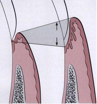
Illustration of pocket formation indicating expansion in two directions (arrows) from the normal gingival sulcus (left) to the periodontal pocket (right)
...

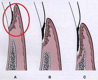
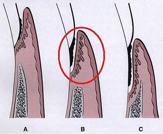
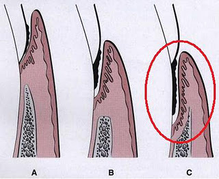
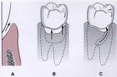
Different types of periodontal pockets. A, Gingival pocket. There is no destruction of the supporting periodontal tissues. B, Suprabony pocket. The base of the pocket is coronal to the level of the underlying bone. Bone loss is horizontal. C, Intrabony pocket. The base of the pocket is apical to the level of the adjacent bone. Bone loss is vertical.
....
Pockets can be classified as follows:
Gingival pocket (pseudo pocket):
This type of pocket is formed by gingival enlargement without destruction of the underlying periodontal tissues. The sulcus is deepened because of the increased bulk of the gingiva
Periodontal pocket:
This type of pocket occurs with destruction of the supporting periodontal tissues. Progressive pocket deepening leads to destruction of the supporting periodontal tissues and loosening and exfoliation of the teeth.
Two types of periodontal pockets exist:
Suprabony (supracrestal or supraalveolar), in which the bottom of the pocket is coronal to the underlying alveolar bone.
Intrabony (infrabony, subcrestal or intraalveolar), in which the bottom of the pocket is apical to the level of the adjacent alveolar bone. In this second type, the lateral pocket wall lies between the tooth surface and the alveolar bone.
Pockets can involve one, two, or more tooth surfaces and can be of different depths and types on different surfaces of the same tooth and on approximating surfaces of the same interdental space . Pockets can also be spiral (i.e., originating on one tooth surface and twisting around the tooth to involve one or more additional surfaces). These types of pockets are most common in furcation areas.
Classification of pockets according to involved tooth surfaces. A, Simple pocket. B, Compound pocket. C, Complex pocket
....
CLINICAL FEATURES
Clinical signs such as bluish-red, thickened marginal gingiva;
A bluish-red vertical zone from the gingival margin to the alveolar mucosa;
Gingival bleeding, suppuration,or both;
Tooth mobility;
And diastema formation and symptoms such as localized pain or pain "deep in the bone" are suggestive of the presence of periodontal pockets.
The only reliable method of locating periodontal pockets and determining their extent is careful probing of the gingival margin along each tooth surface.

A, Extrusion of the central incisor and diastema associated with the periodontal pocket. B, The entire length of the periodontal probe inserted to the base of the periodontal pocket in the central incisor
...
Correlation of Clinical and Histopathologic Features of the Periodontal Pocket
Clinical Features
1. The gingival wall of the periodontal pocket presents various degrees of bluish-red discoloration; flaccidity; a smooth, shiny surface; and pitting on pressure.
2. Less frequently, the gingival wall may be pink and firm.
3. Bleeding is elicited by gently probing the soft tissue wall of the pocket.
4. When explored with a probe, the inner aspect of the periodontal pocket is generally painful.
5. In many cases, pus may be expressed by applying digital pressure.
Histopathologic Features
1. The discoloration is caused by circulatory stagnation; the flaccidity, by destruction of the gingival fibers and surrounding tissues; the smooth, shiny surface, by the atrophy of the epithelium and edema; the pitting on pressure, by edema and degeneration.
2. In such cases, fibrotic changes predominate over exudation and degeneration, particularly in relation to the outer surface of the pocket wall. However, despite the external appearance of health, the inner wall of the pocket invariably presents some degeneration and is often ulcerated.
3. Ease of bleeding results from increased vascularity, thinning and degeneration of the epithelium, and the proximity of the engorged vessels to the inner surface.
4. Pain on tactile stimulation is due to ulceration of the inner aspect of the pocket wall.
5. Pus occurs in pockets with suppurative inflammation of the inner wall.
..
On the basis of depth alone, however, it is sometimes difficult to differentiate between a deep normal sulcus and a shallow periodontal pocket. In such borderline cases, pathologic changes in the gingival distinguish the two conditions.
PATHOGENESIS
The initial lesion in the development ofperiodontitis is the inflammation of the gingiva in response to a bacterial challenge. Changes involved in the transition from the normal gingival sulcus to the pathologic periodontal pocket are associated with different proportions of bacterial cells in dental plaque. Healthy gingiva is associated with few microorganisms, mostly coccoid cells and straight rods. Diseased gingiva is associated with increased numbers of spirochetes and motile rods. However, the microbiota of diseased sites cannot be used as a predictor of future attachment or bone loss because their presence alone is not sufficient for disease to start or progress.
Extension of the junctional epithelium along the root requires the presence of healthy epithelial cells. Marked degeneration or necrosis of the junctional epithelium retards rather than accelerates pocket formation. Degenerative changes seen in the junctional epithelium at the base of periodontal pockets are usually less severe than those in the epithelium of the lateral pocket wall. Because migration of the junctional epithelium requires healthy, viable cells, it is reasonable to assume that the degenerative changes seen in this area occur after the junctional epithelium reaches its position on the cementum.
The transformation of a gingival sulcus into a periodontal pocket creates an area where plaque removal becomes impossible, and the following feedback mechanism is established:
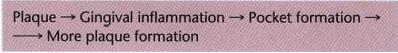
....
The rationale for pocket reduction is based on the need to eliminate areas of plaque accumulation.>>>>>>>>>>>>>... FROM [ندعوك للتسجيل في المنتدى أو التعريف بنفسك لمعاينة هذا الرابط] | |
|